
A. Genovese, V. Piuri, F. Scotti, A Decision Support System for Acute Lymphoblastic Leukemia Detection based on Explainable Artificial Intelligence, Image and Vision Computing, vol. 151, no. 105298, November 2024, ISSN 0262-8856.
Medical imaging
Leukemia Detection
DL4ALL
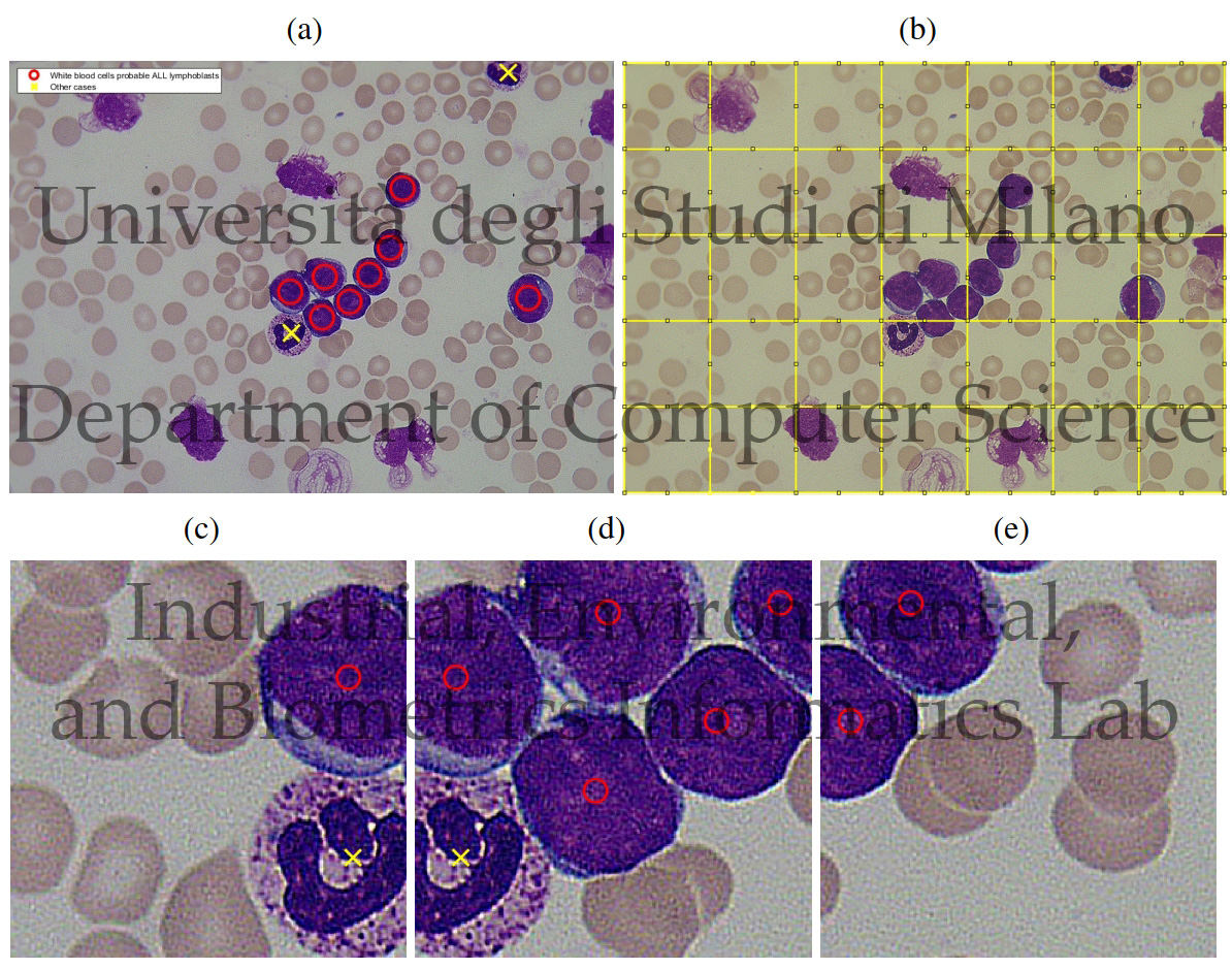
Acute Lymphoblastic (or Lymphocytic) Leukemia (ALL) is a disease that affects the blood cells, can spread rapidly throughout the body, and may result in fatal consequences if not detected at an early stage. One of the techniques routinely used to diagnose ALL consists in analyzing White Blood Cells (WBC) present in peripheral blood samples to look for malformations or abnormalities. Such malformations may be an indicator of lymphoblasts, which naturally occur in the bone marrow. However, an elevated number of WBCs with lymphoblast characteristics may be a sign of ALL.
Traditionally, the analysis of the WBC morphology is performed manually by an expert pathologist, who looks at the blood cells and estimates the concentration of lymphoblasts present in peripheral blood. Such process, being extremely repetitive and time-consuming, may lead to fatigue, with the consequence that the pathologist could miss important information correlated with the presence of ALL.
To overcome the disadvantages of a manual inspection process, Computer Aided Diagnosis (CAD) systems are being increasingly researched: such systems are often based on image processing and machine learning and, by automatically detecting lymphoblasts, can help the pathologist in performing a preliminary screening of the blood samples. Among CAD systems, recent methods are increasingly considering the use of machine learning approaches based on Deep Learning (DL) and Convolutional Neural Networks (CNN), due to their high accuracy in several fields, including medical imaging. In particular, CNNs have the ability of automatically learning data representations, without the need for a handcrafted feature extraction step, with the consequence that CAD systems based on CNNs may be designed with limited knowledge of the application scenario.
Traditionally, the analysis of the WBC morphology is performed manually by an expert pathologist, who looks at the blood cells and estimates the concentration of lymphoblasts present in peripheral blood. Such process, being extremely repetitive and time-consuming, may lead to fatigue, with the consequence that the pathologist could miss important information correlated with the presence of ALL.
To overcome the disadvantages of a manual inspection process, Computer Aided Diagnosis (CAD) systems are being increasingly researched: such systems are often based on image processing and machine learning and, by automatically detecting lymphoblasts, can help the pathologist in performing a preliminary screening of the blood samples. Among CAD systems, recent methods are increasingly considering the use of machine learning approaches based on Deep Learning (DL) and Convolutional Neural Networks (CNN), due to their high accuracy in several fields, including medical imaging. In particular, CNNs have the ability of automatically learning data representations, without the need for a handcrafted feature extraction step, with the consequence that CAD systems based on CNNs may be designed with limited knowledge of the application scenario.
Relevant publications
A. Genovese, V. Piuri, F. Scotti, ALL-IDB Patches: Whole slide imaging for Acute Lymphoblastic Leukemia detection using Deep Learning, Proc. of the IEEE Int. Conf. on Acoustics Speech and Signal Processing Workshops (ICASSPW 2023), pp. 1-5, Rhodes Island, Greece, June 2023, ISSN 979-8-3503-0261-5.
A. Genovese, V. Piuri, K. N. Plataniotis, F. Scotti, DL4ALL: Multi-task cross-dataset transfer learning for Acute Lymphoblastic Leukemia detection, IEEE Access, vol. 11, pp. 65222-65237, 2023, ISSN 2169-3536.
A. Genovese, ALLNet: Acute Lymphoblastic Leukemia detection using lightweight convolutional networks, Proc. of the 2022 IEEE Int. Conf. on Computational Intelligence and Virtual Environments for Measurement Systems and Applications (CIVEMSA 2022), pp. 1-6, Chemnitz, Germany, June 2022, ISSN 978-1-6654-3445-4.
A. Genovese, M. S. Hosseini, V. Piuri, K. N. Plataniotis, F. Scotti, Histopathological transfer learning for Acute Lymphoblastic Leukemia detection, Proc. of the 2021 IEEE Int. Conf. on Computational Intelligence and Virtual Environments for Measurement Systems and Applications (CIVEMSA 2021), pp. 1-6, June 2021, ISSN 978-1-6654-1249-0.
A. Genovese, M. S. Hosseini, V. Piuri, K. N. Plataniotis, F. Scotti, Acute Lymphoblastic Leukemia detection based on adaptive unsharpening and Deep Learning, Proc. of the 2021 IEEE Int. Conf. on Acoustics, Speech, and Signal Processing (ICASSP 2021), pp. 1205-1209, Toronto, ON, Canada, June 2021, ISSN 978-1-7281-7605-5.
R. Donida Labati, V. Piuri, F. Scotti, ALL-IDB: the acute lymphoblastic leukemia image database for image processing, Proc. of the 2011 IEEE Int. Conf. on Image Processing (ICIP 2011), pp. 2045-2048, Brussels, Belgium, September 2011, ISSN 978-1-4577-1302-6.
F. Scotti, Automatic morphological analysis for acute leukemia identification in peripheral blood microscope images, Proc. of the 2005 IEEE Int. Conf. on Computational Intelligence for Measurement Systems and Applications (CIMSA 2005), pp. 96-101, Giardini Naxos - Taormina, Italy, July 2005.
F. Scotti, Robust Segmentation and Measurements Techniques of White Cells in Blood Microscope Images, Proc. of the 2006 IEEE Instrumentation and Measurement Technology Conf. (IMTC 2006), pp. 43-48, Sorrento, Italy, April 2006, ISSN 1091-5281.
V. Piuri, F. Scotti, Morphological classification of blood leucocytes by microscope images, Proc. of the 2004 IEEE Int. Conf. on Computational Intelligence for Measurement Systems and Applications (CIMSA 2004), pp. 103-108, Boston, MA, USA, July 2004, ISSN 0-7803-8341-9.